

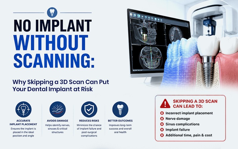
A dental implant should never be placed without a CBCT (Cone Beam CT) 3D scan, especially in patients with minimal bone depth near the nerve canal. Skipping this scan and relying only on 2D X-rays increases the risk of nerve injury during implant placement, which can cause paresthesia — numbness near the lower lip that can be temporary or permanent. At Matrix Dental & Skin Lounge, every implant case is scanned and digitally planned before surgery, under the supervision of Dr. Sourabh Nagpal.
The Problem: Implants Placed Without Proper Scanning
Some dental implants are still placed using only 2D X-rays or chairside radiographs, without a 3D CBCT scan. This is the single biggest risk factor in implant complications involving the nerve canal.
Why 2D X-rays aren’t enough for implant planning:
- A 2D X-ray flattens the three-dimensional jawbone into a single flat image
- It cannot accurately show the depth or exact path of the nerve canal
- It cannot reliably measure how much safe bone exists above the nerve canal
- It leaves the implantologist estimating, rather than measuring, critical distances
Who is most at risk: Patients with minimal bone depth in the lower jaw, where the nerve canal sits close to the area where the implant needs to be placed. In these cases, even a small misjudgment in depth can result in the implant contacting or damaging the nerve.
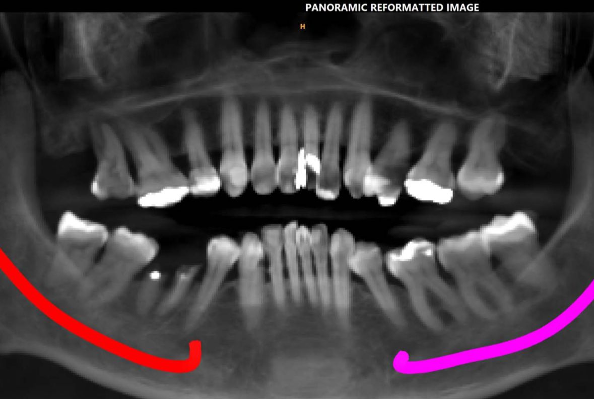
What happens when scanning is skipped or minimal: The implant can be placed too close to, or directly into, the nerve canal. This causes a condition called paresthesia numbness, tingling, or altered sensation, most often felt near the lower lip, chin, or gum on the affected side.
Paresthesia from implant-related nerve injury can be:
- Temporary or permanent
- Short-term or long-term
- Reversible or irreversible
This is why minimal scanning, or no scanning at all, is not a shortcut. It is the leading preventable cause of implant-related nerve complications.
The Solution: A CBCT 3D Scan Before Every Implant
A CBCT (Cone Beam Computed Tomography) scan is a 3D imaging technology that solves the exact problem 2D X-rays create. It gives the implantologist a full three-dimensional view of the jaw, allowing the nerve canal to be located precisely rather than estimated.
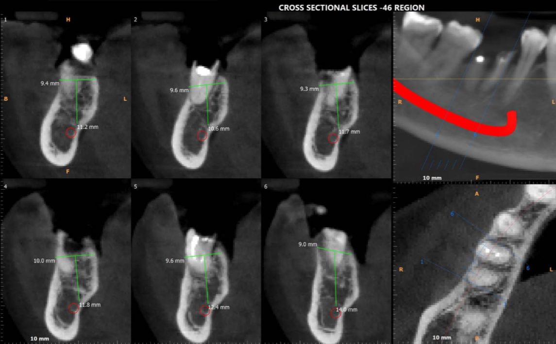
Unlike a standard X-ray, a CBCT scan generates a full 3D image of the jaw. This allows the implantologist to see the exact location, depth, and path of the nerve canal from every angle, rather than relying on a flat projection and clinical guesswork. With this information, the implantologist can:
- Pinpoint precisely how much safe bone depth is available before reaching the nerve canal
- Select an implant size and length that fits safely within that space
- Plan the exact angle and position of placement in advance, rather than adjusting mid-surgery
- Identify cases where the bone depth is too limited for a standard implant, allowing for alternative planning before any drilling begins
In short, a CBCT scan turns implant placement from an educated guess into a precisely guided procedure. The implant size is chosen based on real anatomical measurements, not assumptions, and the nerve canal is treated as a hard boundary that the entire surgical plan is built around, not something to be avoided by chance.
For patients, the rule should be simple: no implant should ever be placed without a proper CBCT scan first. If a clinic is willing to proceed without one, especially in a case with limited bone depth, that is a sign to seek a second opinion.
How Matrix Dental & Skin Lounge Approaches This, Under Dr. Sourabh Nagpal
At Matrix Dental & Skin Lounge, CBCT scanning isn’t an optional add-on offered for complex cases only it’s a non-negotiable first step in the implant journey for every patient. Treatment always begins with a detailed clinical examination followed by a CBCT scan, which gives the implant team a complete 3D map of bone quality, bone volume, and the exact position of the nerve canal before any surgical decision is made.
This evaluation is led by Dr. Sourabh Nagpal, who brings extensive experience in implant dentistry and full-mouth rehabilitation to even the most anatomically challenging cases including patients with minimal bone depth near the nerve canal, where the margin for error is smallest. Using digital implant planning software alongside the CBCT data, Dr. Nagpal and the team determine the ideal implant position, the correct implant size for the available bone, and the safest surgical approach, all before the patient is in the surgical chair.
This is paired with 3D guided implant surgery, which carries the precision from the scan directly into the placement itself, reducing the room for deviation during the procedure. The result is an approach where nerve canal proximity is identified and planned around in advance, not discovered as a complication afterward.
For patients, particularly those who have been told their case is “complicated” due to low bone depth, this distinction matters. A scan-first, plan-first approach doesn’t just improve the odds of a successful implant it’s what stands between a confident, lasting smile and the kind of nerve complication that proper diagnostics exist to prevent.